
What is Coats’ disease?
Coats’ disease is a rare eye condition usually affecting only one eye. The disease is not hereditary, mostly occurring in young males, usually before age 15, but can occur later in life. Symptoms may include a pupil with a white appearance, crossed eyes, or poor visual acuity.
Why is Coats’ disease a problem?
In Coats’ Disease there are changes to the blood vessels in the eye, where the blood vessels leak, causing fluid build-up and leak into the retina (the retina acts like the film in a camera to help us see). This fluid leakage can cause vision loss, and in severe cases, retinal detachment. Coats’ disease can cause blindness if left untreated.
Patients with Coats’ disease can develop glaucoma or retinal detachment, and eventually complete vision loss if the condition is left untreated.
Coats’ disease may also lead to cataract, inflammation, and severe pain in the affected eye.
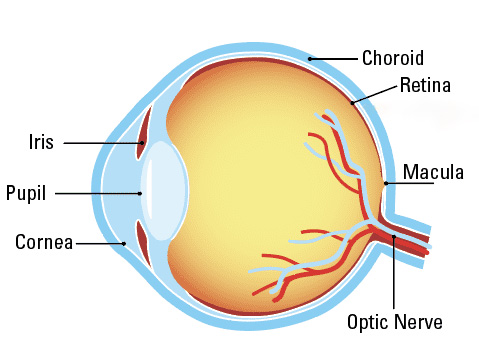
Parts of the Eye
In Coats’ Disease, blood vessels can leak, causing fluid build-up and leaking into the retina.
How is Coats’ disease diagnosed?
Early diagnosis and treatment are key to preventing vision loss from Coats’ disease. An ophthalmologist (an eye doctor with a medical degree: MD or DO) or an optometrist (an eye doctor with an OD degree) will perform a dilated eye exam.
- Dilated eye exam: The eye doctor will dilate (widen) the pupils of the eyes with eye drops to allow a better view of the back of your eyes (retina and macula).
- Fluorescein angiogram: The eye doctor uses a camera to observe your retina and blood vessels in the back of your eye to see if there are any leaking blood vessels.
Let your eye doctor know if you or your child has experienced recent vision changes, symptoms of Coats’ disease.
How is Coats’ disease treated?
Treatment options include:
- Laser: A laser is used to target abnormal blood vessels in the retina.
- Cryotherapy: Abnormal tissue and blood vessels are frozen and destroyed to prevent further leakage.
- Vitreoretinal surgery: Retinal detachment is treated by surgery in later stages of the disease.
- Anti-VEGF therapy: Medications to target vascular endothelial growth factor (VEGF) to reduce abnormal blood vessel growth is used.
- Intravitreal steroids: Steroids are injected in the eye to reduce inflammation and leakage from blood vessels.
- Patching and glasses: To improve the weakened eye, a patch is placed on the unaffected eye to address vision changes.